Home
/ Head And Neck Muscle Diagram : Risorius Png Images Pngwing : Deep muscles of the neck.
Head And Neck Muscle Diagram : Risorius Png Images Pngwing : Deep muscles of the neck.
Head And Neck Muscle Diagram : Risorius Png Images Pngwing : Deep muscles of the neck.. The skull can be further subdivided into. They say nothing that is not said in countless other such diagrams. Obliquus capitis superior also extends from the occiput to c1 while obliquus. § voluntary somatic muscle § make up the skeletal muscles cardiac striated muscle § involuntary visceral muscle § forms most of the walls of the heart and adjacent parts of muscle attachment sites. 9 видео 363 062 просмотра обновлен 5 февр.
Partly act as accessory muscles for respiration. Muscle identification muscle actions and muscle origins and insertions. We hope this picture head and neck muscles diagram can help you study and research. Important exam questions on head & neck. They control heartbeat, breathing, and digestion.
Head And Neck Anatomy Wikipedia from upload.wikimedia.org Muscles allow a person to move, speak, and chew. The head rests on the top part of the vertebral column, with the skull joining at c1. Head and neck anatomy is important when considering pathology affecting the same area. The neck muscles, including the sternocleidomastoid and the trapezius, are responsible for the gross motor movement in the muscular system of the head and neck. At the front, the muscles reach from the jawbone to the sternum and clavicle bones, helping us to move our jaw. Masster, temporalis, medial pterygoid and lateral pterygoid muscles. Rectus capitis posterior major and rectus capitis posterior minor attach the inferior nuchal line of the occiput to the c2 and c1 vertebrae respectively. Muscle identification muscle actions and muscle origins and insertions.
To know the answers, please click on the text highlighted in blue.
Orbital, nasal, oral, auricular and scalp/neck muscles. They control heartbeat, breathing, and digestion. In radiology, the 'head and neck' refers to all the anatomical structures in this region excluding the central nervous system, that is, the brain and spinal cord and their associated vascular structures and encasing. The muscular system consists of various types of muscle that each play a crucial role in the function of the body. They say nothing that is not said in countless other such diagrams. It has two major parts. Many in the neck help to stabilize or move the head. Body spine bifid spinous process carotid tuber. We hope this picture head and neck muscles diagram can help you study and research. Masster, temporalis, medial pterygoid and lateral pterygoid muscles. Learn this topic fast with head and neck muscle anatomy reference charts. Only two of the more obvious and superficial neck. The skull can be further subdivided into.
Discover ideas about muscle diagram. This article describes the anatomy of the head and neck of the human body, including the brain, bones, muscles, blood vessels, nerves, glands, nose, mouth, teeth, tongue, and throat. The suboccipital muscles act to rotate the head and extend the neck. This muscle originates at some of the spinous processes in your thoracic vertebrae (the thoracic vertebrae are the ones where your ribs connect), and it inserts at the transverse processes of the first. 9 видео 363 062 просмотра обновлен 5 февр.
Muscles Of The Head Face And Neck Poster Clinical Charts And Supplies from cdn11.bigcommerce.com Only two of the more obvious and superficial neck. At the front, the muscles reach from the jawbone to the sternum and clavicle bones, helping us to move our jaw. Muscles allow a person to move, speak, and chew. For more anatomy content please follow us and visit our website: Head and neck test questions gross anatomy all cervical vertebra have a: We hope this picture head and neck muscles diagram can help you study and research. Quick run down before we get in the details… this is a superficial muscle that tightens the neck, helps lower the jaw, helps draw down the lower lip and angle of the mouth during grimacing/melancholy expression as it hits the corner of the mouth. Muscle identification muscle actions and muscle origins and insertions.
Human muscle system, the muscles of the human body that work the skeletal system, that are under voluntary control, and that are concerned with movement, posture, and balance.
Important exam questions on head & neck. These muscles are located medially to the ears, superior to the mandible, and inferior to the coronal suture of the skull. Head and neck muscles diagram. Click here to read about mesothelioma and its differential diagnosis and mesothelioma t. The neck muscles move the head in every direction, working in pairs on either side of the body. Orbital, nasal, oral, auricular and scalp/neck muscles. This article describes the anatomy of the head and neck of the human body, including the brain, bones, muscles, blood vessels, nerves, glands, nose, mouth, teeth, tongue, and throat. Masster, temporalis, medial pterygoid and lateral pterygoid muscles. Head and neck muscles labeled anatomical. Rectus capitis posterior major and rectus capitis posterior minor attach the inferior nuchal line of the occiput to the c2 and c1 vertebrae respectively. Head and neck muscles diagram so many muscles that cause migraines arm neck shoulders and back learn all about it at kenhub. Head and neck muscles diagram. The splenius capitis and splenius cervicis, which are located in the back of the neck, work to rotate the head.
For more anatomy content please follow us and visit our website: Click here to read about mesothelioma and its differential diagnosis and mesothelioma t. These muscles are located medially to the ears, superior to the mandible, and inferior to the coronal suture of the skull. Head and neck muscles diagram. It has two major parts.
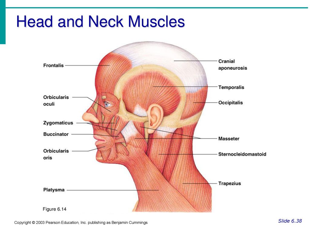
Head And Neck Muscles Figure 6 14 Slide Ppt Download from slideplayer.com Quick run down before we get in the details… this is a superficial muscle that tightens the neck, helps lower the jaw, helps draw down the lower lip and angle of the mouth during grimacing/melancholy expression as it hits the corner of the mouth. Click here to read about mesothelioma and its differential diagnosis and mesothelioma t. Superficial muscles are the muscles closest to the skin surface and can usually be seen while a body is performing actions. Best 25+ neck muscle anatomy ideas on pinterest. Head and neck muscles diagram so many muscles that cause migraines arm neck shoulders and back learn all about it at kenhub. § voluntary somatic muscle § make up the skeletal muscles cardiac striated muscle § involuntary visceral muscle § forms most of the walls of the heart and adjacent parts of muscle attachment sites. They move the head in every direction, pulling the skull and jaw towards the shoulders, spine, and scapula. With one on each side of the neck, these help flex the neck and rotate the head upward and side to side.
Rectus capitis posterior major and rectus capitis posterior minor attach the inferior nuchal line of the occiput to the c2 and c1 vertebrae respectively.
The splenius capitis and splenius cervicis, which are located in the back of the neck, work to rotate the head. Other seemingly unrelated functions, including temperature regulation and vision, also rely. The neck muscles, including the sternocleidomastoid and the trapezius, are responsible for the gross motor movement in the muscular system of the head and neck. The suboccipital muscles act to rotate the head and extend the neck. Mouse over each are to identify the muscle. The neck muscles move the head in every direction, working in pairs on either side of the body. The muscular system consists of various types of muscle that each play a crucial role in the function of the body. The skull can be further subdivided into. Only two of the more obvious and superficial neck. Head and neck muscles labeled anatomical. They control heartbeat, breathing, and digestion. Posted on january 21, 2015 by admin. In radiology, the 'head and neck' refers to all the anatomical structures in this region excluding the central nervous system, that is, the brain and spinal cord and their associated vascular structures and encasing.
Mouse over each are to identify the muscle neck muscle diagram. This article describes the anatomy of the head and neck of the human body, including the brain, bones, muscles, blood vessels, nerves, glands, nose, mouth, teeth, tongue, and throat.
